This is the readme for the models associated with the paper
Wang YJ, Chen BS, Lin MW, Lin AA, Peng H, Sung RJ, Wu
SN. Time-dependent block of ultrarapid-delayed rectifier K(+) currents
by aconitine, a potent cardiotoxin in heart-derived H9c2 myoblasts and
in neonatal rat ventricular myocytes. Toxicol Sci (2008) 106:454-463.
Abstract:
Aconitine (ACO), a highly toxic diterpenoid alkaloid, is recognized to
have effects on cardiac voltage-gated Na(+) channels. However, it
remains unknown whether it has any effects on K(+) currents. The
effects of ACO on ion currents in differentiated clonal cardiac (H9c2)
cells and in cultured neonatal rat ventricular myocytes were
investigated in this study. In H9c2 cells, ACO suppressed
ultrarapid-delayed rectifier K(+) current (I(Kur)) in a time- and
concentration-dependent fashion. The IC(50) value for ACO-induced
inhibition of I(Kur) was 1.4 microM. ACO could accelerate the
inactivation of I(Kur) with no change in the activation time constant
of this current. Steady-state inactivation curve of I(Kur) during
exposure to ACO could be demonstrated. Recovery from block by ACO was
fitted by a single-exponential function. The inhibition of I(Kur) by
ACO could still be observed in H9c2 cells preincubated with ruthenium
red (30 microM). Intracellular dialysis with ACO (30 microM) had no
effects on I(Kur). I(Kur) elicited by simulated action potential (AP)
waveforms was sensitive to block by ACO. Single-cell Ca(2+) imaging
revealed that ACO (10 microM) alone did not affect intracellular
Ca(2+) in H9c2 cells. In cultured neonatal rat ventricular myocytes,
ACO also blocked I(Kur) and prolonged AP along with appearance of
early afterdepolarizations. Multielectrode recordings on neonatal rat
ventricular tissues also suggested that ACO-induced
electrocardiographic changes could be associated with inhibition of
I(Kur). This study provides the evidence that ACO can produce a
depressant action on I(Kur) in cardiac myocytes.
--------------------------
To run the model:
XPP: start with the command
xpp ode\RatAPtest01.ode
or xpp RatAPtest01.ode
Mouse click on Initialconds, and then (G)o.
This makes a trace similar to the upper part of fig 6A, 6C and 7 in
the paper of Wang et al.
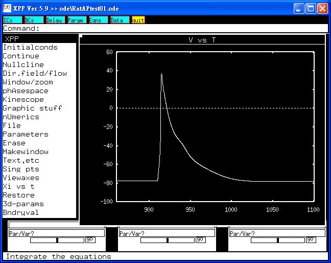 This trace was used as a template of rat ventricular myocyte and
replayed to H9c2 myoblasts in an attempt to evoke I(Kur).
Bard Ermentrout's website http://www.pitt.edu/~phase/
describes how to get and use xpp (Bard wrote xpp).
The model file was submitted by:
Dr. Sheng-Nan Wu
Department of Physiology
Natl Cheng Kung U Med Coll
Tainan 70101, Taiwan
This trace was used as a template of rat ventricular myocyte and
replayed to H9c2 myoblasts in an attempt to evoke I(Kur).
Bard Ermentrout's website http://www.pitt.edu/~phase/
describes how to get and use xpp (Bard wrote xpp).
The model file was submitted by:
Dr. Sheng-Nan Wu
Department of Physiology
Natl Cheng Kung U Med Coll
Tainan 70101, Taiwan