This is the readme for the model associated with the paper:
Lopez-Jury L, Meza RC, Brown MTC, Henny P, Canavier CC (2018)
Morphological and biophysical determinants of the intracellular and
extracellular waveforms in nigral dopaminergic neurons: A
computational study. J Neurosci
http://dx.doi.org/10.1523/JNEUROSCI.0651-18.2018
This model was contributed by L Lopez-Jury and requires NEURON which is freely available from https://www.neuron.yale.edu
Usage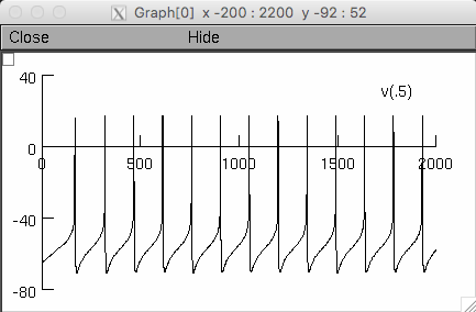
 The above, if you zoom in, is similar to those in Figure 3D
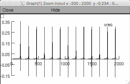
Some notes
about the extracellular simulations, a ShapePlot with the neuron
morphology and two windows showing the somatic intracellular membrane
potential (top) and the extracellular potential recorded close to the
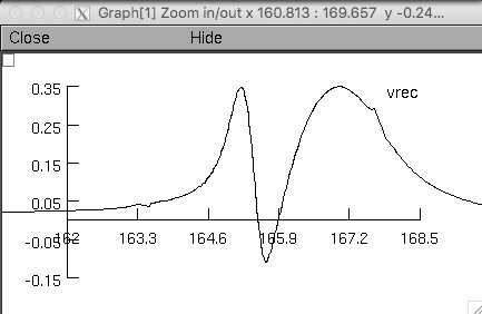
soma (bottom). You may zoom-in to note the waveform of the
extracellular spike.
--
To check how AIS length modify the shape of the action potential use
"L" NEURON parameter by typing: ais.L Original reconstruction length
is 41.45 microns, try changing it to 10 or 100 microns to see the
effect on the waveform. From our results, the range of AIS length of
16 dopaminergic neuron reconstructions is from 19 to 58 microns.
--
To better visualize the effect of the AIS length on the intracellular
shape of action potential you may calculate the first derivative of
membrane potential or generate a phase plane plot of the spike. The
effect on the extracellular waveform is clearly visible.
--
To change from 2-domain model to 3-domain model, open "parameters.hoc"
file and change the sodium and delayed rectifier potassium conductance
densities to 200 and 100 respectively. As indicated in the
comments. This will change the conductance only in the soma section,
dendrites keep their conductances fix.
--
Also, you may change the neuron 3D reconstruction by changing the
second line in "parameters.hoc" file. We have included two different
reconstructions: one from a mouse and the other from a rat. Both
neuron models reproduce the extracellular waveform obtained by
experimental recordings without changing any parameter.