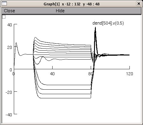
This is the readme for this model that tries to mimic the VS model
illustrated in the papers by Borst and Haag (1996,1997, et al. 1999)

1. Borst A, Haag J (1996) The intrinsic electrophysiological
characteristics of fly lobula plate tangential cells: I. Passive
membrane properties. J Comput Neurosci 3:313-36
2. Haag J, Vermeulen A, Borst A (1999) The intrinsic
electrophysiological characteristics of fly lobula plate tangential
cells: III. Visual response properties. J Comput Neurosci 7:213-34
3. Haag J, Borst A (1997) Encoding of visual motion information and
reliability in spiking and graded potential neurons. J Neurosci
17:4809-19

Executing this model will produce a figure similar to fig 10 from Haag
1997.


Created by B. Torben-Nielsen (TENU @ OIST) and Ted Carnevale
2009-01-20

There are key unresolved differences between the published model and
the NEURON implementation.

The published model's damped oscillations occur when the low- to
moderate- amplitude depolarizing currents are injected; in the NEURON
implementation, they are triggered by low-amplitude hyperpolarizing
currents.

The published model responds fairley linearly to a wide range of
hyperpolarizing currents; the NEURON implementation shows a quite
nonlinear jump to negative membrane potentials when medium to large
hyperpolarizing currents are injected.

The published model's resting potential is 0; the resting potential of
the NEURON implementation is nonzero and nonuniform. The NEURON
implementation's resting potential can be forced to zero everywhere by
adjusting e_pas in each compartment so that i_pas cancels out the
contributions of local Na and K currents; however, the resulting
model's responses to injected depolarizing and hyperpolarizing
currents diverge even more from those of the published model.

Moreover,there is still an unresolved issue concerning the time
constants.  According to Borst, these are not scaled/normalized in
figure 8 (f,g) from the 1997 paper. This implies that our time
constants are tau_max times to large.

When contacted, Borst was positive about the constructed model.  The
discrepancies are likely due to (1) the morphology and (2) the
distribution of the voltage-gated channels.  Recall that (i) the
morphology is coming from neuromorpho.org; a curated database in which
they "repair" morphologies.  A different morphology leads to a
different outcome (especially since the tiny branches are close to the
axon).  (ii) the description of the location of the voltage-gated
channels is rather vague.  Clearly this may effect the outcome as
well.  (Qualitatively the results are similar, quantitatively, there
are some discrepancies).