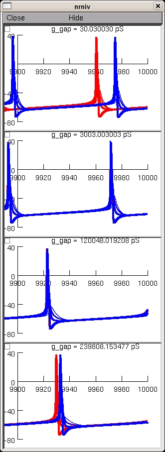
This is the readme for the model for the paper:

Saraga F, Ng L, Skinner FK (2006) Distal gap junctions and active
dendrites can tune network dynamics. J Neurophysiol 95:1669-82

These model files were supplied by Dr Saraga.

cella.hoc: Morphology of cell a
cellb.hoc: Morphology of cell b (same as cell a)
gap.mod: gap junction defined as a resistor
gapdist.hoc: connects the 2 cells at ~400 microns from soma
gapdist.ses: opens graphs to display GJ current and soma voltage for both cells
gapmid.hoc: connects the 2 cells at ~200 microns from soma
gapmid.ses: opens graphs to display GJ current and soma voltage for both cells
gapprox.hoc: connects the 2 cells at ~100 microns from soma
gapprox.ses: opens graphs to display GJ current and soma voltage for both cells
hh.mod: Hodgkin-Huxley sodium and potassium currents
initdist: Opens all files for distal gap junctions
initmid: Opens all files for middle gap junctions
initprox: Opens all files for proximal gap junctions
prog.hoc: Inserts HH currents into cell a and cell b

Usage:

The runs typically take tens of minutes so these simulations are best
run with batch scripts.  To recreate a figure similar to fig 6 in the
paper you can autolaunch from modeldb or download and extract the
archive then under

mswin
-----

Compile the mod files with mknrndll and then double click on the
mosinit.hoc

unix/linux
----------

Compile the mod files nrnivmodl then start the simulation with
nrngui mosinit.hoc

MAC OS X
--------

Drag and drop the folder created in the extraction onto the mknrndll
icon.  Drag and drop the mosinit.hoc file onto the nrngui icon.

----------------

Once the simulation is started you can explore the model with the
graphical interface (see esp. the miscellaneous functions provided by
extras.hoc) or press the create fig 6 button which will generate a
panel similar to fig 6.  This takes tens of minutes to complete.  It
should generate a figure like this: