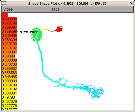
These NEURON files are for the model in the paper:

Gouwens, NW and Wilson, RI (2009)
Signal propagation in Drosophila central neurons.
J Neurosci 29(19):6239-6249.

Abstract:

Drosophila is an important model organism for investigating neural
development, neural morphology, neurophysiology, and neural correlates
of behaviors. However, almost nothing is known about how electrical
signals propagate in Drosophila neurons. Here we address these issues
in antennal lobe projection neurons (PNs), one of the most
well-studied classes of Drosophila neurons. We use morphological and
electrophysiological data to deduce the passive membrane properties of
these neurons and to build a compartmental model of their electrotonic
structure. We find that these neurons are electrotonically extensive,
and that a somatic recording electrode can only imperfectly control
the voltage in the rest of the cell. Simulations predict that action
potentials initiate at a location distant from the soma, in the
proximal portion of the axon. Simulated synaptic input to a single
dendritic branch propagates poorly to the rest of the cell and cannot
match the size of real unitary synaptic events, but we can obtain a
good fit to data when we model unitary input synapses as dozens of re-
lease sites distributed across many dendritic branches. We also show
that the true resting potential of these neurons is more
hyperpolarized than previously thought, due to the experimental error
introduced by the electrode seal conductance. A leak sodium
conductance also contributes to the resting potential. Taken together,
these findings have fundamental implications for how these neurons
integrate their synaptic inputs. Our results also have important
consequences for the design and interpretation of experiments aimed
at understanding Drosophila neurons and neural circuits.

To run:

Remember to first compile the .mod files for your system:

mswin:
------
Run mknrndll from the NEURON program group.
Double click the mosinit.hoc file or one of the hoc files beginning
with "figure..."

Mac OS X:
---------
drag and drop the folder (dm1_pn_model) onto the mknrndll icon.  Drag
and drop the mosinit.hoc file onto the nrngui icon.

unix/linux:
-----------
run "nrnivmodl" in the folder (dm1_pn_model), then "nrngui mosinit.hoc".

After the simulation is started it will automatically generate plots
similar to those in the paper. The neuronal morphologies are defined
in the files dm1_morph_1.hoc, dm1_morph_2.hoc, and dm1_morph_3.hoc.
For example running figure_4a_cell1.hoc (or pressing the cell 1 button
after auto-launching from ModelDB or running mosinit.hoc) should lead
to a figure like: